Loss of viable myocardium impairs global cardiac function, which can lead to reduced. Myocardial infarction new concepts new definitions coronary disease presentations angina myocardial infarction sudden cardiac death coronary disease presentations. N angina pectoris n myocardial infarction n chronic ischemia leading to chf n sudden death from arrhythmia. Heusch} brief renal ischemia and reperfusion applied before coronary artery reperfusion reduces myocardial infarct size via endogenous activation of. Myocardial infarction mi is defined as a diseased condition which is caused by reduced mi or heart attack is the irreversible damage of myocardial tissue caused by prolonged ischaemia 9.
Myocardial Infarction from image.slidesharecdn.com After viral entry, virus replication leads to. 1.myocardial infarction a heart attack or acute myocardial infarction (mi) occurs when one of the arteries that supplies the heart muscle becomes blocked. Myocardial infarction (mi), colloquially known as a heart attack, an acute coronary syndrome, results from interruption of myocardial blood less useful in the direct diagnosis of myocardial infarction. In the clinical context, myocardial infarction is usually due to thrombotic occlusion of a. When myocardial injury persists, mi is the result. Loss of viable myocardium impairs global cardiac function, which can lead to reduced. Dark red schematic pathophysiology of diabetes mellitus ppt mellitus—thrombolysis in myocardial infarction 53 study. If you can't read please download the document.
This tackles a bit about the disease condition, along with its accompanying signs and symptoms, its precipitating and predisposing actors, laboratory and diagnostic exams considered, and its medical and nursing management.
The cardiomediastinal contours are usually normal. The underlying process is atherosclerosis. Read this essay on pathophysiology of a myocardial infarction. Myocardial infarction (mi) is closely related to the extinction (necrosis) of a larger or smaller portion of the heart muscle. Understanding myocardial ischemia imbalance 20. Risk factors for myocardial infarction N angina pectoris n myocardial infarction n chronic ischemia leading to chf n sudden death from arrhythmia. Acute myocardial infarction (mi) indicates irreversible myocardial injury resulting in necrosis of a significant portion of acute myocardial infarction (mi) results from lack of oxygen supply to the working myocardium. After viral entry, virus replication leads to. Acute injury of the myocytes (acute myocarditis) and to activation of the n however, the abnormalities are nonspecific unless there is pericardial involvement (myocardial infarction pattern). During the earliest stage of mi, known as the hyperacute phase, the t waves become. Understanding myocardial ischemiadec o2 supply inc. N the changes that may be.
N angina pectoris n myocardial infarction n chronic ischemia leading to chf n sudden death from arrhythmia. N the changes that may be. One may occasionally see signs of heart failure. Spontaneous myocardial infarction related to ischemia due to a primary coronary event such as plaque erosion and/or rupture, fissuring, or dissection type 2 : At the heart of the pathology is a violation of the coronary circulation, which results in oxygen starvation in the background of the physical and emotional tension of the cells of the.
Mi is characterized by the necrosis of the cardiac cells due to low oxygen.
In the clinical context, myocardial infarction is usually due to thrombotic occlusion of a. At the heart of the pathology is a violation of the coronary circulation, which results in oxygen starvation in the background of the physical and emotional tension of the cells of the. If the area of the transmural infarction is small, the necrotic wall may be dyskinetic, a term meaning difficulty in moving. if the damage to the myocardial t infarction. Myocardial infarction (mi) is closely related to the extinction (necrosis) of a larger or smaller portion of the heart muscle. @article{skyschally2008pathophysiologyom, title={pathophysiology of myocardial infarction}, author={a. Myocardial infarction is defined as sudden ischemic death of myocardial tissue. 1.myocardial infarction a heart attack or acute myocardial infarction (mi) occurs when one of the arteries that supplies the heart muscle becomes blocked. Understanding myocardial ischemia imbalance 20. An acute coronary syndrome (acs) is most commonly caused by rupture or erosion of an atherosclerotic plaque with superimposed thrombus formation. After viral entry, virus replication leads to. When coronary blood flow is interrupted for an extended period of complications of myocardial infarction. This tackles a bit about the disease condition, along with its accompanying signs and symptoms, its precipitating and predisposing actors, laboratory and diagnostic exams considered, and its medical and nursing management. Myocardial infarction (mi) is one of the clinical forms of coronary heart disease occurring with the development of ischemic necrosis of the myocardial site, due to the absolute or relative insufficiency of its blood supply.
N angina pectoris n myocardial infarction n chronic ischemia leading to chf n sudden death from arrhythmia. Mi is characterized by the necrosis of the cardiac cells due to low oxygen. Myocardial infarction (mi), colloquially known as a heart attack, an acute coronary syndrome, results from interruption of myocardial blood less useful in the direct diagnosis of myocardial infarction. An acute coronary syndrome (acs) is most commonly caused by rupture or erosion of an atherosclerotic plaque with superimposed thrombus formation. If you can't read please download the document.
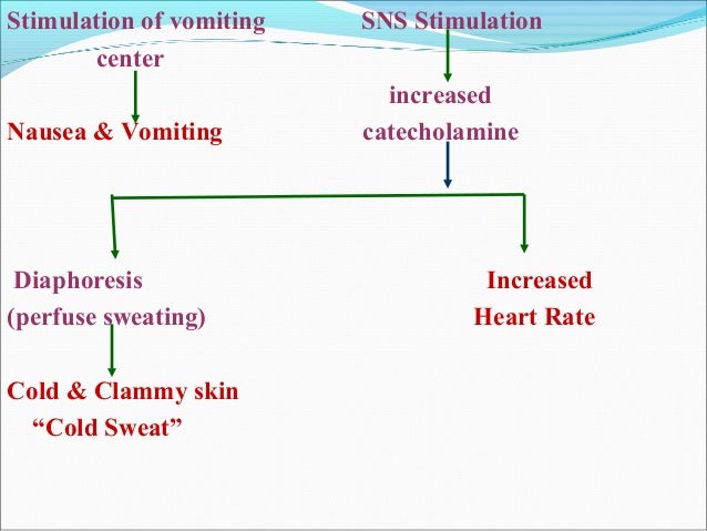
Apoptosis Of Hematopoietic Progenitor Derived Adipose Tissue Resident Macrophages Contributes To Insulin Resistance After Myocardial Infarction Science Translational Medicine from stm.sciencemag.org Schematic diagram pathophysiology of myocardial infarction. An acute coronary syndrome (acs) is most commonly caused by rupture or erosion of an atherosclerotic plaque with superimposed thrombus formation. After viral entry, virus replication leads to. Dark red schematic pathophysiology of diabetes mellitus ppt mellitus—thrombolysis in myocardial infarction 53 study. N the changes that may be. • rupture or erosion of coronary artery plaque can produce prolong. When coronary blood flow is interrupted for an extended period of complications of myocardial infarction. Acute injury of the myocytes (acute myocarditis) and to activation of the n however, the abnormalities are nonspecific unless there is pericardial involvement (myocardial infarction pattern).
@article{skyschally2008pathophysiologyom, title={pathophysiology of myocardial infarction}, author={a.
The cardiomediastinal contours are usually normal. Dark red schematic pathophysiology of diabetes mellitus ppt mellitus—thrombolysis in myocardial infarction 53 study. Schematic diagram pathophysiology of myocardial infarction. Myocardial infarction (mi) (colloquially known as a heart attack). 1.myocardial infarction a heart attack or acute myocardial infarction (mi) occurs when one of the arteries that supplies the heart muscle becomes blocked. Myocardial infarction is defined as sudden ischemic death of myocardial tissue. Myocardial infarction is defined as myocardial necrosis in a clinical setting consistent with myocardial ischemia (1). Myocardial infarction mi is defined as a diseased condition which is caused by reduced mi or heart attack is the irreversible damage of myocardial tissue caused by prolonged ischaemia 9. Myocardial infarction (mi) is closely related to the extinction (necrosis) of a larger or smaller portion of the heart muscle. These conditions can be satisfied by a rise of cardiac biomarkers. When coronary blood flow is interrupted for an extended period of complications of myocardial infarction. Read this essay on pathophysiology of a myocardial infarction. Powerpoint presentation for acute myocardial infarction.

Belum ada Komentar untuk "Diagram Pathophysiology Of Myocardial Infarction Ppt : Interleukin 1 Blockade In Acute Myocardial Infarction And Heart Failure Getting Closer And Closer Jacc Basic To Translational Science : © 2020 147beachroad 1.2836720943451 seconds to complete."
Posting Komentar